
In the search for personalized epilepsy treatment: physicist Tena Dubček’s new approach to medicine

What Epilepsy seizures can look like! Source: The Experimentalist
Aleksandra Nelson
Epileptic seizures can feel dramatically different from one person to another. Having epilepsy means that the neurons in your brain can suddenly go crazy and begin to fire strong signals uncontrollably to their neighbors, causing a seizure. For different people, seizures happen in different regions of the brain, so they experience very different effects. With such complexity and diversity of symptoms, it is impossible to find a single treatment that will help everyone.
Dr. Tena Dubček, a senior researcher in the Department of Health Sciences and Technology at ETH Zurich, is working toward a personalized approach to understanding and treating epilepsy.
The stakes are high. Epilepsy affects 50–70 million people worldwide, making it one of the most common neurological disorders. Despite advances in treatment, nearly one-third of patients continue to experience uncontrolled seizures. People with epilepsy remain in constant fear as, at any moment, they could lose consciousness and fall, be it in the kitchen or at the top of the stairs. Sometimes the last resort for these people is to target the brain directly, either through surgery or electric stimulation. These methods have a long history and have improved many lives. However, as Dr. Dubček explains, they often rely on generalizations about the brain and fail to account for individual differences among patients.
We are all used to personalized recommendations in our everyday lives. Music streaming platforms analyze our listening history and suggest new songs and artists that we might like. What if the same principle could be applied to treating diseases, particularly epilepsy? In today's era of data-driven medicine, researchers can collect individual patients' brain maps and analyze them to identify the most effective treatment plan for each person.
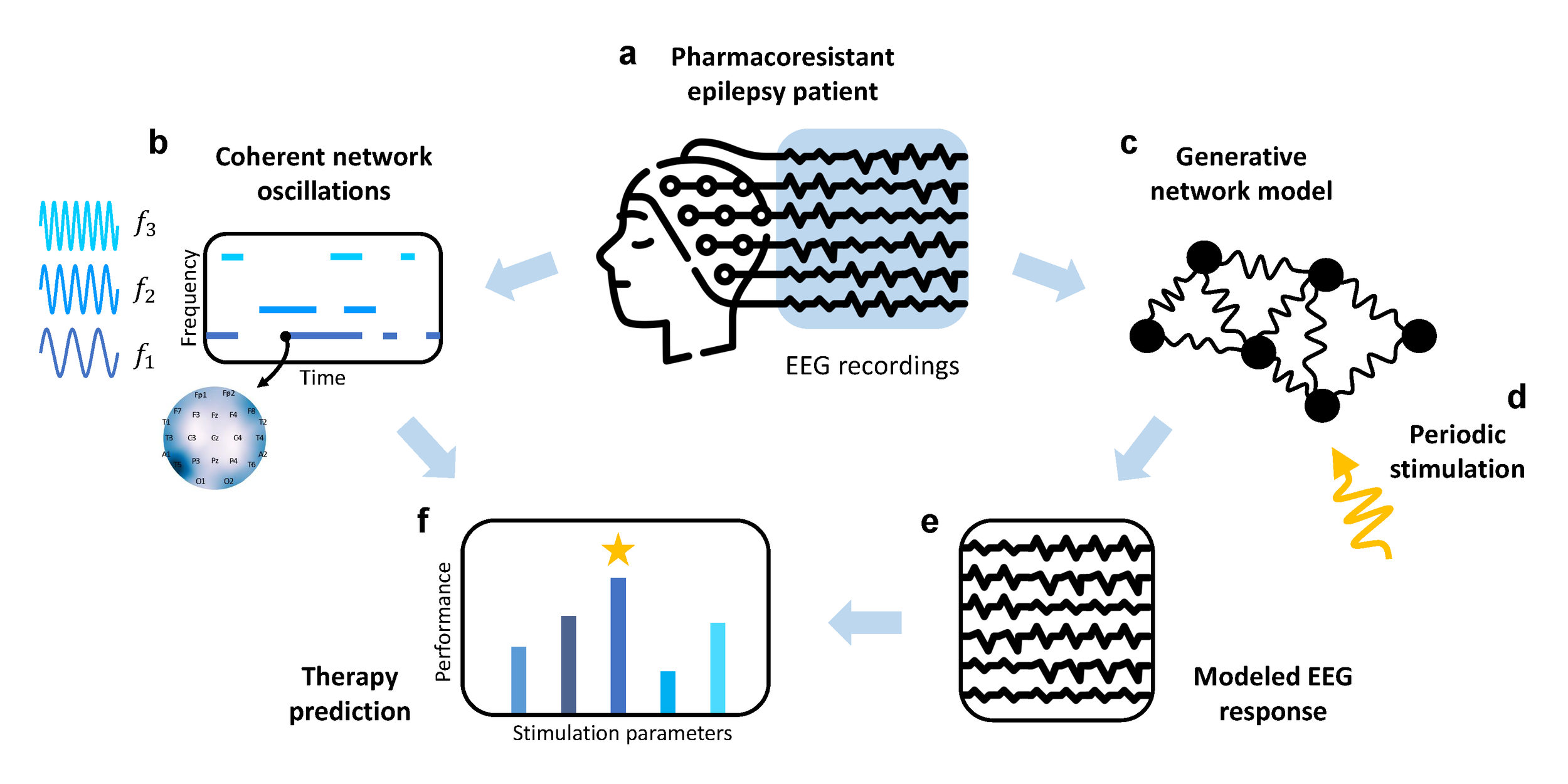
Identifying unique brain oscillations in epilepsy patients and using computational models to develop personalized brain stimulation therapies. Adapted from Clinical Neurophysiology (2025)
The first step is already in place: when visiting a neurologist, a patient usually undergoes an electroencephalogram(EEG), which records the brain's electrical activity. The doctor places many small electrodes all around the patient’s head and measures their electrical signals for 20-30 minutes. This procedure gives a lot of information about the patient’s brain, what regions show the strongest activity, and how this activity changes and oscillates over time.
EEG is crucial for diagnosing epilepsy. Doctors usually look for patterns directly visible to the naked eye, like spikes and slower-than-usual rhythms. In many cases, these patterns are enough to diagnose and classify epilepsy. But this is like skimming a synopsis of a crime story instead of reading the whole book — you know who the killer is, but you miss a lot of the details and reasoning. EEG is a rich source of information, so it makes sense to use it as a tool and develop new methods to extract more detailed insights from brain activity.
This was Dr. Dubček’s goal when she joined Professors Dr. Rafael Polania and Dr. Lukas Imbach at ETH Zurich. “I personally related to epilepsy. It surprised me how rarely quantitative methods are used in clinics, at least compared to my own research experience. So, as a physicist with a strong machine-learning background, I moved into epileptology to change this,” she explains.
Dr. Dubček soon realized that clinicians work with real people suffering from real diseases, so they are very careful in incorporating new methods, like machine learning. For doctors, results must be interpretable. Many machine learning methods that are so useful in other areas, from self-driving cars to discovering new materials, are not ideal for clinical research. While they could take EEG as an input and output a treatment recommendation, this would essentially be a black box. Doctors wouldn't understand the reasoning behind the recommendation, so it wouldn't be safe for patients. Learning more about doctors’ needs, Dr. Dubček became a bridge between the worlds of quantitative and clinical research.
Dr. Dubček had a challenging goal: to analyze EEG data in a quantitative and patient-specific way, and make sure that her methods remain clinically interpretable. As a physicist, she is used to seeing an object of study as a system of interacting parts, like atoms and electrons in a material. The way these parts interact and affect each other determines how the object behaves as a whole. Dr. Dubček was therefore surprised to discover that this idea is quite new in clinical brain research. Usually, doctors analyze the activity of different brain regions independently, and only recently have they begun to view the brain as a network whose components influence each other. Dr. Dubček is convinced that analyzing the brain as a complex network will bring new insights into epilepsy research.
A similar approach was taken by researchers at Johns Hopkins University, led by Prof. Dr. Sridevi V. Sarma, in a study published in Nature Neuroscience. By treating the EEG as a network, the researchers analyzed how bursts of activity in one brain region were followed by activity in connected regions moments later. For each patient, they identified the regions that are most likely to cause strong oscillations in the rest of the brain — they termed these the focal point for epileptic seizures. This way, doctors can precisely determine which parts of the brain should be acted upon during surgery.
Instead of looking at immediate connections between regions, Dr. Dubček had another idea in her work, published in 2025 in Clinical Neurophysiology. She analyzed the entire EEG to identify long-lasting brain oscillations that do not change or disappear. She found that such stable oscillations differ between healthy individuals and patients experiencing a prolonged and severe seizure known as status epilepticus. Dr. Dubček emphasizes, “Our method analyzes the brain as a whole network, allowing us to see how different regions are connected and where these oscillations occur.” This makes the discovered oscillations visual and easily interpretable by doctors, who know how different oscillations correspond to a patient’s cognitive state. In the future, this method will help doctors understand the differences between epileptic and healthy brain activity.
But this is just the first step. By focusing on the most stable oscillations, Dr. Dubček can create a computer model of the brain for each individual patient. She can then test how the model reacts to external stimuli and find a stimulus that will return it from a status epilepticus to a healthy state. This stimulus will be unique to each patient and can be used as a personalized treatment to control their seizures.
However, one step is still missing to make this work, and this is the subject of Dr. Dubček’s future research. “We know how stimulation works on single neurons. But we don’t really know how to map the dynamics of single neurons onto the EEG. There is so much missing in this area,” she explains. This knowledge gap appears at the intersection of two fields: in clinics, doctors usually talk about EEG, while neuroscientists talk in terms of single neurons. Connecting two different fields of science is a challenging task, but it can open entirely new ways of understanding this complex disease.